Precursor of breast cancer or not?
New mouse model bridges 10-20 years of development of DCIS into breast cancer within one year
An international team of researchers led by the Netherlands Cancer Institute and in collaboration with Colinda Scheele (VIB-KU Leuven Center for Cancer Biology) developed a method to better predict the outgrowth of Ductal Carcinoma In Situ (DCIS), a possible precursor of breast cancer, into invasive breast cancer. Using mice into which cells from women with DCIS were inserted, researchers can better identify which DCIS patients are at risk for breast cancer.
The study was published in the scientific journal Cancer Cell and is part of PRECISION, a major international research project on DCIS funded by Cancer Research UK and the Dutch Cancer Society as part of Cancer Grand Challenges – a global funding initiative co-founded by Cancer Research UK and the National Cancer Institute in the US.
DCIS consists of aberrant cells in the milk ducts of the breast. In the Netherlands, it is found in about 2,300 women a year, about 80% of them detected at breast cancer screening. This is because calcium splashes can be seen on the X-ray of the breast (the mammogram), which may indicate DCIS.
Preventing overtreatment
To this day, however, it is not possible to predict which DCIS will progress to breast cancer and which will not. Therefore, virtually all women with DCIS are treated preventively through mastectomy or breast-conserving surgery followed by radiation and, in some countries, hormone therapy. As a result, tens of thousands of women around the world undergo intensive treatment without any benefit, but suffer the disadvantages.
To prevent overtreatment in the future, researchers at The Netherlands Cancer Institute in collaboration with Colinda Scheele of the VIB-KU Leuven Center for Cancer Biology developed a "living biobank" of DCIS cells to better understand their progression to cancer.
Following the “live” process from DCIS to breast cancer
The development from DCIS to breast cancer in humans usually takes 10 to 20 years. In the new mouse models, in which human DCIS cells are inserted, this takes only one year. Also, it is difficult to study DCIS in patients themselves because DCIS is surgically removed fairly soon after diagnosis.
Researchers at the Netherlands Cancer Institute developed more than 100 different DCIS mouse models that allow them to accurately follow the process from DCIS to breast cancer. DCIS cells from surgical tissue were introduced into the milk ducts of mice under anesthesia, using a very thin needle. The growth of these very different DCIS abnormalities was then followed over a period of one year, with just under half of the mice developing invasive breast tumors. Thanks to this time saving, this study allowed scientists to better understand why DCIS does or, more importantly, does not grow into breast cancer.
Research leader Jos Jonkers of the Netherlands Cancer Institute: "Thanks to our mouse models, we were able to study DCIS live for the first time. This yielded a wealth of information. We hereby bridge a gap of several decades between preclinical research and the clinic."
Molecular similarities between humans and mice
While transferring tissue from women with DCIS to mice was quite an achievement in itself, the results of the study are even more interesting. Thanks to the new mouse models, researchers have also gained important information about breast cancer risk factors in DCIS. For example, molecular studies showed that the presence of the HER2 protein increases the risk of breast cancer. In contrast, presence of the protein to which the hormone estrogen can bind, known as hormone-sensitive breast cancer, meant a lower risk.
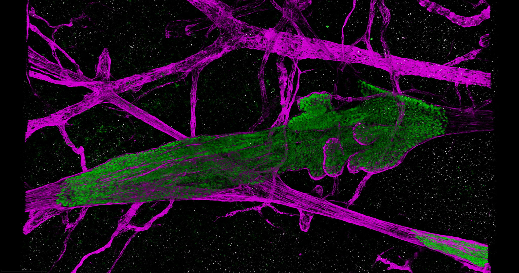
3D Microscopy detects different growth patterns
After a year, half of the mouse models with DCIS were found not to have developed breast cancer while the other half did. To study what factors had led to this distinction, the mouse models were also examined with three-dimensional microscopy by the research group of Colinda Scheele of the VIB-KU Leuven Center for Cancer Biology. The group developed a method for mapping tissue cell-by-cell with this. For example, the research group discovered that human DCIS cells exhibit two different three-dimensional growth patterns that can predict breast cancer risk. For example, they found that in most mice where breast cancer did not develop, the DCIS cells had replaced the mouse cells in the milk ducts ("replacement growth"), while if breast cancer did develop, the milk ducts were "inflated" by the DCIS cells ("expansive growth").
Colinda Scheele, VIB-KU Leuven Center for Cancer Biology: “Thanks to the good work of our colleagues at the NKI, we were able to examine more than 700 samples. That we were able to discover from the growth pattern which DCIS populations respectively do or do not lead to breast cancer is promising. If in the future we can also demonstrate in human patients which DCIS cells will not evolve into cancer, we can potentially save them a lot of physical and mental suffering.”

Better distinguish between harmless and high-risk DCIS
The mouse models have provided a better understanding of the biology of DCIS. This information may help to better distinguish in the future between DCIS abnormalities that are very likely to be harmless and those DCIS abnormalities that may well grow into breast cancer.
“In the first case, we could then consider not treating these women and only screening them regularly. In the second case, treatment is probably necessary to protect these women from breast cancer in time,” says pathologist Jelle Wesseling of the Netherlands Cancer Institute.
Wesseling is team lead for the Cancer Grand Challenges PRECISION consortium funded by the Dutch Cancer Society and Cancer Research UK, in which researchers from the Netherlands, England and America are researching the risk of DCIS developing into invasive breast cancer. The current research with mouse models is also part of this consortium.
Unique biobank collection for science
Some of the mouse models now form a unique biobank and can be used by scientists around the world to gather new information. The DCIS cells from these models can be frozen after outgrowth in the mouse and later reintroduced into mice. Even after three generations of mice, the molecular properties of the DCIS cells appear to be remarkably stable. PhD student Stefan Hutten, who conducted the research, said: "We now have a unique collection of nineteen distributable models representing all molecular subtypes of DCIS."
Publication: Stefan Hutten et al, ' A living biobank of patient-derived ductal carcinoma in situ Mouse-INtraDuctal xenografts identifies risk factors for invasive progression', Cancer Cell, April 27, 2023. DOI:


Joran Lauwers
About Cancer Grand Challenges
Co-founded in 2020 by two of the largest funders of cancer research in the world: Cancer Research UK and the National Cancer Institute, Cancer Grand Challenges supports a global community of diverse, world-class research teams to come together, think differently and take on some of cancer’s toughest challenges. These are the obstacles that continue to impede progress and no one scientist, institution or country will be able to solve them alone. With awards of up to $25M, Cancer Grand Challenges teams are empowered to rise above the traditional boundaries of geography and discipline to make the progress against cancer we urgently need.
About the VIB-KU Leuven Center for Cancer Biology
Cancer has many causes. Often it is a combination of lifestyle, environmental factors and genetic variation. We need to fight cancer on many fronts, and this can only be done by using knowledge. The VIB-KU Leuven Center for Cancer Biology researchers unravel new mechanisms in order to develop both specific diagnostic methods and treatments.